BIO INX Material Enables Functional Cartilage Regeneration in Rabbit Study
BIO INX Material Enables Functional Cartilage Regeneration in Rabbit Study
Zwijnaarde, Belgium 27/08/2026 BIO INX is proud to announce that its biodegradable high-resolution printing resin, DEGRAD INX, has been successfully used in a groundbreaking preclinical study demonstrating the regeneration of functional cartilage in rabbits. The research, conducted by scientists at TU Wien in the research group of Prof. Aleksandr Ovsianikov, by PhD Student Oliver Kopinski- Grünwald and collaborators from the Ludwig Boltzmann Institute for Traumatology; AUVA and the Medical University of Vienna; all within the Austrian Cluster for Tissue Regeneration, was recently published in the European Polymer Journal and represents an important milestone in the field of regenerative medicine.
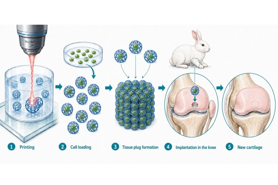
From 3D printing to new Cartilage: the building block is printed using multiphoton lithography, filled with cells, combined into a plug, allowed to mature and placed inside the knee of the rabbit and grows there into new tissue.
The study describes an innovative approach in which cell-laden implantable microscaffolds of around 0.3mm diameter were produced using BIO INX’ DEGRAD INX resin through high resolution laser-based 3D printing also known as multiphoton lithography. These intricate 3D printed microstructures were loaded with stem-cell-derived cartilage cell spheres were cultured and assembled into tissue constructs capable of regenerating osteochondral critical-sized defects in a rabbit model.
Using these biodegradable microscaffolds, the researchers of TU WIEN created tissue building blocks that protected and organized the cells for implantation. The approach holds great promise for as a novel tissue engineering strategy, where the scaffolds could be injected to fill irregular cartilage defects in a minimally invasive manner. Once implanted, the constructs promoted the formation of healthy cartilage tissue that integrated well with the surrounding native tissue.
After twelve weeks, the treated defects demonstrated highly encouraging results. Animals receiving the DEGRAD INX-based implants showed significantly improved cartilage regeneration compared to untreated controls, with the regenerated tissue covering a substantially larger portion of the defect and reaching approximately 91% of the thickness of native cartilage. Histological analysis further classified the repaired tissue as "nearly normal cartilage," highlighting the strong regenerative potential of the technology.
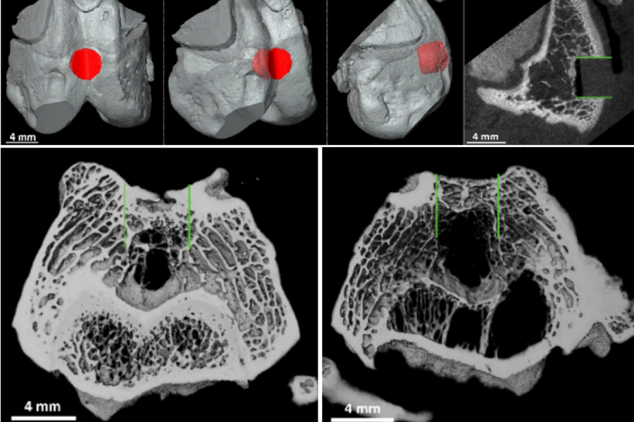
Top: 3D image of the introduced defect and µCT image of the defect before treatment.
Bottom Left: µCT image of a knee joint treated with the cell containing 3D printed microscaffolds. Bottom right: the control group (right), where the green lines indicate the diameter and depth of the created defect. A clear improvement is observed in the treated group. (Image Courtesy of TUWIEN)
It is the first publication showing this technology in living animals, what’s more the study indicated very promising results showing active cartilage regeneration in the treated defects. This is of significant importance, as cartilage is a tissue with very poor regenerative ability. “The technology of microscaffolds for minimally invasive tissue engineering is a strategy that we have been working on for the past 10 years, but having the first succesful in vivo results are very encouraging that we are on the right track to translate this towards the clinics” said Prof. Aleksandr Ovsianikov of TUWien.
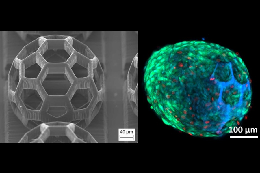
Left: Electron microscopy image of a printed microscaffold; Right: Confocal microscopy image of the stem cells (green) inside the 3D printed cage in DEGRAD INX (blue) (Image courtesy of TUWIEN)
Multiphoton lithography is one of the most advanced additive manufacturing technologies available today, enabling the fabrication of complex three-dimensional structures with micrometer and even sub-micrometer resolution. This unprecedented resolution allows researchers to create highly sophisticated cellular microenvironments that were previously impossible to manufacture.
A key enabler of this work is DEGRAD INX, BIO INX's biodegradable and biocompatible resin specifically developed for multiphoton lithography. The material uniquely combines sub-micrometer printing resolution with full biodegradability and biocompatibility, allowing printed structures to provide temporary support during tissue formation before gradually degrading as new tissue develops. This combination addresses one of the major challenges in tissue engineering.
The study demonstrates that combining high-resolution biodegradable biomaterials with advanced biofabrication techniques can enable minimally invasive regenerative therapies for cartilage repair, one of the most challenging tissues in the human body to regenerate due to its limited natural healing capacity.
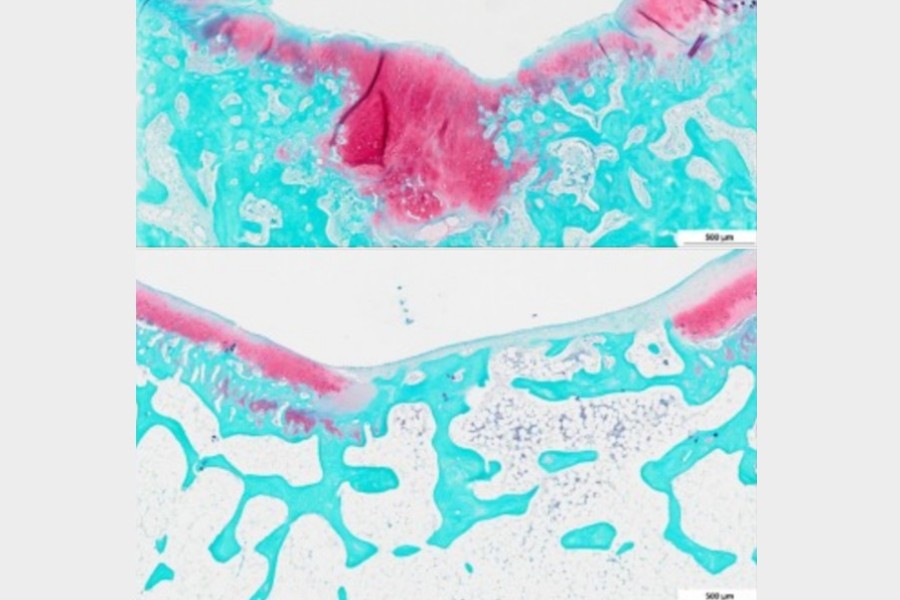
Top: Histology image showing newly formed cartilage (pink) at the tissue defect versus the control group without the printed structures but bone ingrowth from below (bottom) indicating cartilage formation. (Image Courtesy of TUWien)
"This publication represents another important validation of our DEGRAD INX platform," said Aysu Arslan, CSO and Co-founder of BIO INX. "By combining the unmatched resolution of multiphoton lithography with a fully biodegradable material, researchers can fabricate sophisticated cell carriers that simply were not possible before. Seeing these materials contribute to the regeneration of cartilage in vivo is an exciting step towards future regenerative therapies for patients."
Although further research and clinical validation as well as translation are still required, these promising preclinical results represent an important stepping stone toward future minimally invasive treatments for cartilage injuries and, ultimately, human cartilage regeneration.
The full study can be found here: https://doi.org/10.1016/j.eurpolymj.2026.114891